
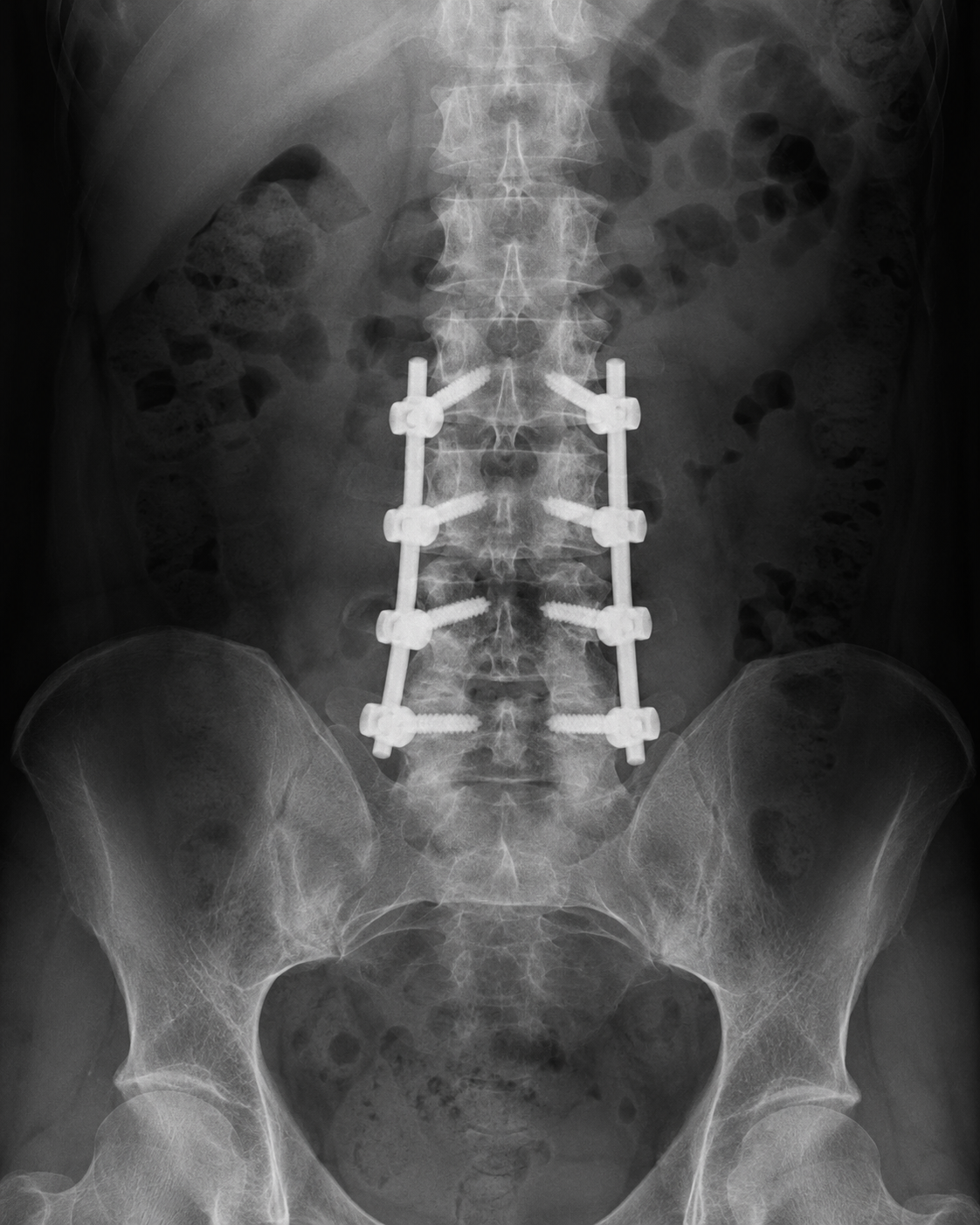
Lumbar Fusion Surgery in Los Angeles
Orthopedic Spine Surgeon — Long Beach & Torrance, CA
Medically reviewed by Brandon A. Ortega, MD · Last updated July 30, 2026
Lumbar fusion surgery permanently joins two or more vertebrae in the lower back to treat instability, deformity, or painful abnormal motion. Selected procedures can be performed using minimally invasive techniques that reduce muscle disruption and incision size. Many patients gradually return to light daily activities within several weeks, although the vertebrae may require six months to a year to form a mature fusion. Reduced nerve-related pain, improved stability and mobility, maintained spinal alignment, and progressive bone growth on follow-up imaging are common indicators of success.
What Is Lumbar Fusion Surgery?
Lumbar fusion is a spinal stabilization procedure that connects two or more vertebrae in the lower back. Bone-graft material is placed between or alongside the vertebrae so that new bone gradually develops across the treated level.
Screws, rods, cages, or other implants are commonly used to hold the vertebrae in alignment while the fusion develops. The hardware provides immediate structural support, but the biological fusion process occurs gradually over several months.
Fusion eliminates movement between the treated vertebrae. This may reduce pain caused by instability or abnormal motion, prevent further vertebral slippage, correct deformity, or maintain stability after compressed nerves have been surgically decompressed.
Lumbar fusion may be performed through several approaches:
- Anterior lumbar interbody fusion (ALIF): The disc is reached through the abdomen.
- Posterior lumbar interbody fusion (PLIF): The disc space is approached from the back.
- Transforaminal lumbar interbody fusion (TLIF): The disc is approached from one side of the back.
- Lateral lumbar interbody fusion: The disc space is reached through the patient’s side.
- Posterolateral fusion: Bone graft is placed along the back and sides of the vertebrae.
Some procedures use minimally invasive incisions, muscle dilators, tubular retractors, navigation, and image-guided instrumentation. A traditional open approach may be more appropriate when extensive decompression, multilevel reconstruction, or significant deformity correction is required.
Conditions Treated
Lumbar fusion may be considered for lower-back conditions involving instability, deformity, abnormal movement, or nerve compression, including:
- Lumbar spondylolisthesis
- Degenerative disc disease
- Lumbar spinal instability
- Lumbar spinal stenosis with instability
- Degenerative scoliosis
- Recurrent disc herniation
- Severe facet-joint degeneration
- Vertebral fractures
- Lumbar deformity or loss of normal alignment
- Instability following laminectomy or another decompression
- Failed previous lumbar surgery
- Selected spinal tumors or infections
- Nerve-root compression associated with an unstable spinal segment
Spondylolisthesis occurs when one vertebra slips forward or backward relative to the adjacent vertebra. When the slippage produces instability or nerve compression, decompression and fusion may be used to stabilize the affected segment.
Lumbar spinal stenosis does not always require fusion. Decompression alone may be sufficient when the spine is stable. Fusion may be added when there is existing instability, vertebral slippage, significant deformity, or concern that decompression would destabilize the treated level.
Fusion may also be used after a damaged disc is removed. A cage or spacer restores height between the vertebrae and provides an area where the bone graft can develop into a solid fusion.
Lumbar fusion is not an appropriate treatment for every case of lower-back pain. Outcomes are generally more predictable when imaging, examination findings, and symptoms identify a specific structural problem that fusion can address.
Surgery Candidates and General Patient Criteria
Many patients begin with nonsurgical care before lumbar fusion is considered. Treatment may include medication, activity modification, physical therapy, spinal injections, weight management, or temporary bracing.
Potential candidates may have:
- Persistent symptoms despite appropriate conservative care
- Confirmed lumbar instability or vertebral slippage
- Lower-back pain associated with abnormal spinal movement
- Leg pain, numbness, or weakness caused by nerve compression
- Difficulty standing, walking, working, or completing daily activities
- Spinal deformity requiring stabilization or correction
- Imaging findings that correspond with the patient’s symptoms
- Adequate bone quality to support the implants and fusion
- General health suitable for anesthesia and surgery
- Realistic expectations regarding recovery and symptom improvement
Surgery may be considered sooner when there is progressive weakness, severe instability, an unstable fracture, worsening neurological function, or another condition that threatens the nerves or spinal structure.
Bone quality is especially important because the vertebrae must support screws, cages, and other implants while the fusion heals. Osteoporosis or osteopenia may increase the risk of implant loosening, vertebral fracture, cage subsidence, or failure of the bones to fuse.
A bone-density assessment may therefore be recommended, particularly for older adults or patients with risk factors for weakened bone.
Nicotine use also interferes with bone and wound healing. Patients may be required to stop smoking, vaping, or using nicotine products before surgery and remain nicotine-free throughout recovery.
Other factors that may affect candidacy or healing include:
- Diabetes
- Obesity
- Poor nutrition
- Active infection
- Certain autoimmune or inflammatory conditions
- Long-term steroid use
- Previous lumbar surgery
- The number of levels requiring fusion
- The patient’s ability to follow postoperative restrictions
The surgical evaluation may include a physical and neurological examination, standing X-rays, bending or extension X-rays, MRI, CT imaging, and bone-density testing. An abnormal scan alone does not establish the need for fusion; the findings should correspond with the patient’s symptoms and examination.
What to Expect
Before Surgery
Before lumbar fusion, the surgical team reviews the patient’s symptoms, medical history, previous treatments, medications, allergies, and diagnostic imaging.
Preoperative preparation may include:
- Physical and neurological examination
- Updated X-rays, MRI, or CT imaging
- Blood and urine testing
- Electrocardiogram or other medical testing
- Bone-density assessment
- Primary-care or specialist clearance
- Review of medications and supplements
- Instructions regarding blood-thinning medication
- Nicotine cessation
- Fasting before anesthesia
- Skin preparation
- Arranging transportation and help at home
Patients should tell the surgical team about all prescription medications, over-the-counter products, supplements, allergies, bleeding conditions, previous anesthesia reactions, and nicotine use. Medication should not be stopped unless directed by the treating clinician.
The surgeon will explain the proposed surgical approach, the levels being fused, the planned implants and bone graft, the expected hospital stay, activity restrictions, and reasonable treatment alternatives.
Patients may also be encouraged to prepare their home for recovery by placing commonly used items within easy reach and arranging assistance with meals, transportation, household tasks, and childcare.
During Surgery
Lumbar fusion is generally performed under general anesthesia. The patient’s position depends on whether the spine will be approached from the front, side, or back.
Although the precise operation varies, the surgeon generally:
- Makes one or more incisions to access the lumbar spine.
- Confirms the correct spinal level using imaging.
- Removes damaged disc material, bone, or ligament when required.
- Decompresses affected spinal nerves when necessary.
- Prepares the vertebral surfaces for fusion.
- Places bone graft between or alongside the vertebrae.
- Inserts a cage or spacer into the disc space when performing an interbody fusion.
- Places screws, rods, plates, or other instrumentation to stabilize the spine.
- Confirms implant placement and spinal alignment.
- Closes the incision and applies a dressing.
Minimally invasive lumbar fusion uses smaller access points and specialized instruments to reach the spine while limiting disruption to surrounding muscles. These techniques may reduce blood loss, postoperative discomfort, and recovery time for properly selected patients. They do not change the need for the vertebrae to heal together after surgery.
The length of surgery depends on the approach, number of levels, need for nerve decompression, previous surgery, and complexity of the reconstruction.
Patients undergoing a limited fusion may leave the hospital sooner than those having open or multilevel surgery. For example, Cleveland Clinic notes that hospitalization after TLIF may last approximately three to four days, although actual discharge timing varies considerably.
Recovery and Rehabilitation
Patients are usually encouraged to stand and begin walking shortly after surgery with assistance. Walking supports circulation, helps restore mobility, and reduces some complications associated with prolonged bed rest.
Early recovery instructions may include:
- Taking short, frequent walks
- Keeping the incision clean and dry
- Taking medication as directed
- Avoiding heavy lifting
- Limiting bending and twisting
- Avoiding strenuous or high-impact exercise
- Wearing a lumbar brace when prescribed
- Remaining nicotine-free
- Attending postoperative appointments
- Completing follow-up imaging
- Beginning physical therapy when approved
Pain around the incision and stiffness in the lower back are expected during the early recovery period. Leg pain caused by direct nerve compression may improve relatively quickly, but numbness, tingling, or weakness can take longer because nerves recover gradually.
Patients may resume many basic daily activities within several weeks. Return to work may range from a few weeks to several months depending on the procedure, number of levels fused, symptoms, and physical requirements of the job.
A patient with a sedentary position will generally return sooner than someone whose work involves heavy lifting, climbing, repetitive bending, prolonged driving, or operating machinery.
Physical therapy may be introduced as healing progresses. Rehabilitation may address:
- Walking endurance
- Core and lower-body strength
- Posture
- Balance
- Hip mobility
- Safe lifting mechanics
- Gradual return to work or exercise
The patient may feel substantially better before the vertebrae have fully fused. Activity restrictions should therefore be based on the surgeon’s clinical and imaging assessments rather than pain levels alone.
Recovery after ALIF, PLIF, TLIF, or another lumbar fusion often continues for several months. Complete recovery may take up to a year as bone graft develops across the treated level.
Potential Complications and Success Indicators
Potential complications of lumbar fusion include:
- Infection
- Poor wound healing
- Bleeding
- Blood clots
- Reaction to anesthesia
- Nerve-root injury
- Dural tear and spinal-fluid leakage
- New or worsening weakness or numbness
- Persistent or recurrent pain
- Failure of the vertebrae to fuse
- Hardware loosening, movement, or breakage
- Cage or implant subsidence
- Loss of spinal alignment
- Vertebral fracture
- Pain at a bone-graft donor site
- Degeneration of nearby spinal levels
- The need for revision surgery
Different surgical approaches carry additional risks. An anterior approach involves structures within the abdomen, including major blood vessels, while lateral approaches may affect nerves near the hip or thigh. Posterior surgery may involve greater disruption of the lower-back muscles, depending on the technique used.
Failure of the bones to heal into a solid fusion is called nonunion or pseudarthrosis. Factors that may increase the risk include nicotine use, poor bone quality, diabetes, poor nutrition, multilevel fusion, and excessive stress on the spine during recovery.
Common indicators of a successful outcome include:
- Reduced pain traveling into the buttock or leg
- Improved lower-back stability
- Increased walking or standing tolerance
- Improved strength or sensation
- Better ability to work, sleep, or perform daily activities
- Maintained or improved spinal alignment
- Stable screws, rods, and cages on imaging
- Progressive bone growth across the fused level
- No abnormal movement between the treated vertebrae
- Reduced dependence on pain medication
Fusion is generally more reliable for correcting instability, vertebral slippage, deformity, or another clearly identified structural problem than for treating generalized lower-back pain without a confirmed source.
A successful fusion does not guarantee complete pain relief, restore a damaged nerve immediately, or prevent degeneration at other spinal levels.
Patients should promptly contact their surgical team for fever, increasing redness or wound drainage, uncontrolled pain, new or worsening weakness, loss of sensation, difficulty urinating, new bladder or bowel dysfunction, chest pain, or shortness of breath.
Lumbar Fusion Surgery in Los Angeles Q&A
Is lumbar fusion surgery minimally invasive?
Some lumbar fusion procedures can be performed using minimally invasive techniques. The surgeon may use smaller incisions, muscle dilators, tubular retractors, and image-guided instrumentation. Complex deformities, multilevel disease, extensive scar tissue, or major reconstruction may require an open approach.
Does lumbar fusion eliminate all movement in the lower back?
No. The procedure eliminates movement only between the vertebrae included in the fusion. The remaining lumbar and pelvic joints continue to move. The effect on overall flexibility depends on the location and number of levels fused.
How does the surgeon determine whether the fusion has healed?
Healing is evaluated through symptoms, physical examination, and follow-up imaging. X-rays or CT scans may show new bone developing across the treated level, stable hardware, maintained alignment, and an absence of abnormal movement between the fused vertebrae.
Why Choose BAO Spine?
Fellowship-trained expertise
Advanced, subspecialty training in complex and minimally invasive spine surgery.
Conservative care first
Most patients improve without surgery — treatment begins with the least invasive options.
Motion-preserving techniques
Cervical and lumbar disc replacement that protects your natural range of motion.
Two convenient locations
Personalized spine care in both Long Beach and Torrance, California.

About Dr. Ortega
Brandon A. Ortega, MD, is a fellowship-trained orthopedic spine surgeon providing comprehensive, personalized spinal care to adults in Long Beach and Torrance, California. His dedication to helping each patient regain mobility, reduce pain, and improve their quality of life has earned the respect of everyone in the community struggling with neck and back pain.
Consultation
Get expert care with lumbar fusion surgery in Los Angeles
Request an appointment and Brandon A. Ortega, MD's team will reach out to schedule your visit — fellowship-trained, personalized spine care.
Prefer to call? Contact us.